1.1 Mass Spectrometry

Mass Spectrometry of peptide: PolyR

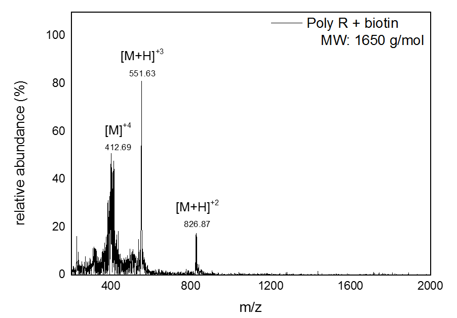
The results seen above shows the ESI-MS spectrum of conjugated Biotin to via PolyR. Since molecular ions are formed in the sample volume after being ionized through the ESI-MS, this helps researchers predict where their theoretical mass/charge ratio based on the relative abundancy as viewed in peaks. Given that the total Molecular Weight for conjugated Biotin to PolyR is 1650 g/mol, it can be safely assumed that the peak will be viewed 825 m/z. We can visibly observe this assumption to hold true, which indicate our sequence specificity was correct and successful. Other peaks that shows less mass than the molecular ion are the result of fragmentation of the molecule. This makes sense since not all solid phase peptides are properly synthesized.

Mass Spectrometry of peptide: Transportan

ESI-MS spectrum of Biotin to Transportan. The total Molecular Weight for conjugated Biotin to Transportan is 3067 g/mol. Thus, a peak should be found at 1533.5 m/z. Many reaction pathways can occur for fragmentation, but only newly formed cations will show up in the mass spectrum, not radical fragments or neutral fragments.

1.2 Dynamic Light Scattering

DLS of LUVs before bursting

In order to confirm that the liposomes were synthesized and to characterize their size, Dynamic Light Scattering (DLS) was performed. DLS is a very sensitive technique with ability to detect particles with diameters as low as 0.3 nm. It measures Brownian motion to determine the size of particles suspended in a liquid.[ 1, 2 ]

The graph above represents the acquired data for dialysed liposomes (26.7 nM) prepared using a polycarbonate membrane with a pore size of 200 nm. The sharp peak around 164.18 nm indicates that the size of the particles is very consistent and sample preparation was very good. The sample yield was also relatively high indicated by the 13.1 % intensity.

DLS of LUVs after bursting

DLS determined to be a good method to verify the bursting of liposomes upon the addition of cell-bursting peptides. This was verified by analyzing the intensity curve and shift in the size of the liposomes. The absence of meaningful data from the Poly-R peptides prompted us to focus our attention on the more robust Trans peptides. The graph above is the result of addition of 4000 nM Trans peptides conjugated to 200 nM quantum dots in 1:1 ratio with the liposomes.

An estimated 8-10 potential streptavidin binding sites are present on one QD. We observed that bursting of liposomes only occurs when most of the binding sites on the QD are occupied. A 20-fold excess of Trans peptides was added to ensure binding on all sites. The sudden presence of much smaller particles (37.84 nm) in addition to the presence of very large particles (5.6 μm) indicates the breakage of many liposomes. This is very likely to result in aggregation of lipids due to the strong hydrophobic interactions from the fatty acid tails. Coupled with a significant decrease in the intensity of the presence of liposomes of the same size, from 13.1% to 8.4%, is another very strong indication of bursting having taken place.

1.3 UV-Vis

UV-Vis: undialysed liposomes

For further verification of bursting, liposomes encapsulating single-stranded DNA were prepared. The remaining ssDNA in the buffer solution was filtered out with rotary filtration dialysis to avoid interference in Ultraviolet-visible spectroscopy (UV/Vis) measurements

UV/Vis was performed using a Nanodrop in order to quantify the concentration of the encapsulated DNA released from the liposomes due to bursting. In this experiment, we used two controls: the undialyzed and the dialyzed liposomes. The undialyzed, unfiltered liposomes contained 9.99 nM of ssDNA in the buffer solution. This concentration decreased to 6.16 nM after dialysis, indicating that dialysis had a positive effect.

UV-Vis: dialysed liposomes

Filtered liposomes were also used in the bursting experiments. The sample containing a 1:1 ratio of QD/peptides to dialyzed liposomes demonstrated an increase in the concentration of DNA to 10.81 nM in the buffer solution, which was even higher than the unfiltered liposomes. This provides further verification that bursting did in fact take place. The ssDNA concentration increased to 12.13 nM in sample with ratio 1:2, which was almost double the concentration of the filtered liposomes. This differs from the DLS data, which suggested that the optimum bursting took place at the 1:1 ratio. However, since the concentrations are quite small, this discrepancy may not be significant. For the last sample, the concentration of ssDNA is decreased to 6.50 nM, a value closer to the concentration of the DNA in the buffer of the filtered liposomes. Similar to the DLS results, this suggests that the increased volume of liposomes for a ratio of 1:10 QD/peptide to liposomes has a severe hindrance on the effect and efficiency of the peptides.

1.4 Gel Electrophoresis

Amicon purification of 6HB

A 10-fold excess of the staples for the 6HB was purified with amicon filtration. Following the annealing reaction, amicon filtration of 6HB was successful in removing the excess staple strands as seen in the second lane. By cutting out and extracting the specific bands from the gel, purified 6HBs can be obtained but at low concentrations.

Magnetic bead purification of 6HB conjugated with streptavidin

After amicon filtration, the 6HB were functionalized with streptavidin and purified with magnetic beads. For verification of remaining 6-helix bundles (6HB) in our solution after purification and modification, a 0.75% agarose gel was performed. Lane A shows the amicon purified 6HB, and lane B indicates streptavidinylated handles at slightly lower concentration. The presence of a very strong signal of a lighter fragment below the signal containing the 6HB indicates the presence of a significant concentration of 'release oligonucleotides' used for strand displacement from the magnetic beads.

Cryo-TEM and WF

Cryo-TEM

Unilamellar liposomes were obtained from the synthesis of 200 nm liposomes(LUVs). The use of this technique allowed us to view our LUVs as single particles allowing us to characterise them based on structure and shape

Wide-field Microscopy

Wide field microscopy was used to capture GUVs stained with diI. GUVs as big as 90 μM were observed at a magnification to 40x with emission of QD at 570 μM. Concentration of GUVs from electroporation were estimated to be ~50 μM.

2.3 Confocal Microscopy

Confocal of GUVs before bursting

GUVs prepared with DiI staining were imaged as a control for bursting experiments. Confocal microscopy was used to capture Biotinylated-GUVs that were conjugated with Streptavidin-Biotin DiI complexes as illuminated above. Likewise, with Wide Field Microscopy: GUVs were formed as big as 90μM were observed. Images were taken before QD-conjugating peptides were added. A large number of GUVs are visibly seen above.

Confocal after bursting GUVs

Bursting of most of the GUVs was observed on the addition of 4000 nM QD + 800 nM Trans peptides. When compared after the addition of QD-conjugating Peptides, there was a rapid and significant decrease of GUVs during confocal microscopy as seen above.

2.4 STORM

STORM

Super-resolution microscopy (STORM) was conducted on 200 nm LUVs and 200nm LUVs containing Poly-R cell-bursting peptides conjugated to quantum dots. The image containing only liposomes is a sample frame from several seconds of data collection. Overall, this sample contains more particles than the sample containing the peptides, which could be an indication of bursting. Supplemented with other bursting data, in can be said that there is a uniform distribution of bursting upon the addition of cell-bursting peptides.

STORM

According to the sigma values obtained from the Gaussian curve, the sample mixed with peptides achieved more precise measurements, indicated by a mean sigma value of 0.0149. The average sigma value of just the liposomes was 0.02388. This decrease in measurement precision could be explained by the presence of more particles in sample with liposomes only. Overall, the sample containing the peptides in addition to the liposomes had smaller particles, suggesting that the peptides may have caused breakage of the liposomes, which then reformed into smaller, less uniform liposomes. Compariing these data to the data obtained from confocal imaging with the GUVs, there was a similar outcome in the reduction of liposomes seen after the addition of peptides.

SEM and TEM

SEM

SEM image of the 6HB functionalised with streptavidin handle sequences.

TEM

TEM image of amicon purified 6HB diluted to 2 nM

2.7 Phase Contrast Microscopy

Controlled Motion of Dynabeads

Linear assembly of Dynabeads of 2.7 μm diameter under the influence of a magnetic fields of 6 mT.

Controlled Motion of Dynabeads

Rotational motion of Dynabeads of 2.7 μm diameter. The velocity observed is 114 rad/s

3 Modeling and Simulation

Qualitative picture of the simulation.

The slice plots show the magnetic flux density. The arrows indicate the magnetic field (H) strength and direction. In the middle part of coils magnetic field is uniformly distributed and has the same vector. The change in the current intensity (AC mode) will result in the reorientation of the magnetic field and therefore in the rotational movement of Dynabeads

Y-X, Z-Y planes of the two Helmholtz coil system

The arrows in the center of the coil system indicate uniformly distributed magnetic flux density which is the result of stationary time simulation. The winding results in a uniform magnetic field between the coils with the primary component parallel to the axis of the two coils. The uniform field is the result of the sum of the two field components parallel to the axis of the coils and the difference between the components perpendicular to the same axis.

  1. Goldburg, W. I. 'Dynamic Light Scattering', American Journal of Physics, vol. 67/no. 12, pp. 1152-1160.(1999)
  2. Malvern Instruments. Dynamic Light Scattering: an introduction in 30 min